The Diethylstilbestrol Legacy : A Powerful Case Against Intervention in Uncomplicated Pregnancy

image of DES drugs

Rebecca Troisi, ScD, Elizabeth E. Hatch, PhD, and Linda Titus, PhD. Pediatrics, 2016

Although the basic tenet of medicine is “First, do no harm,” history is filled with good intentions that were at best unhelpful and at worst harmful. Because medicine seeks to cure afflictions, there is an overwhelming desire on the part of health providers and patients to administer treatment. In certain settings, treatment can be reasonable despite a risk of adverse consequences: for example, if the disease is cured or its morbidity abated and the treatment consequences are less disabling than the disease itself.

In the absence of overt disease, the question of whether to apply an intervention is far more challenging. The safety of interventions must be weighed against the population’s level of risk, the morbidity and/or mortality associated with the disease, and the intervention’s efficacy (eg, BRCA1 mutation, mastectomy, reduced breast cancer risk). Interventions must meet an especially high standard of safety and efficacy when administered in low-risk populations or in settings in which the morbidity associated with the disease is minor. In the worst-case scenario, an intervention may be both ineffective for its primary purpose and cause iatrogenic illness.

Interventions in pregnancy are especially problematic because of the complex physiology of the condition and the possibility of causing short- and long-term adverse consequences in both the mother and her offspring. The continuing story of diethylstilbestrol (DES), a synthetic estrogen, shows the importance of caution when evaluating the merits of interventions involving pregnant women. With regard to DES, investigators believed that pregnancy loss was caused in part by a decrease in estrogen and that administering DES to pregnant women would help maintain a healthy pregnancy. Moreover, because endogenous estrogen concentrations increase dramatically during a healthy pregnancy, supplementation with DES was deemed harmless. During its early years of use, DES was administered to women with threatened pregnancy loss or a history of pregnancy loss. Eventually, DES was advertised to the medical community for “routine prophylaxis in ALL pregnancies” and administered to women with otherwise healthy pregnancies.

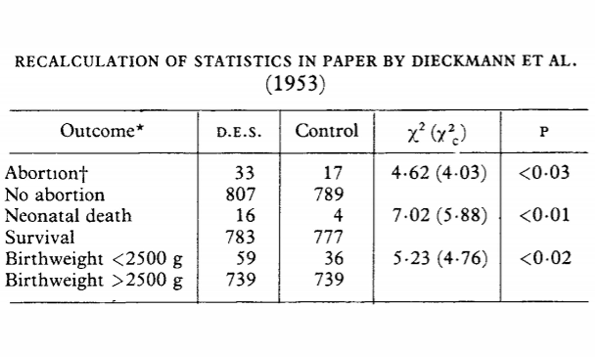
By the time DES was formally evaluated, it was standard of care in high-risk obstetrics practices. The first clinical trial to determine the efficacy of DES, reported in 1953, showed that DES did not improve pregnancy outcome. (Indeed, a subsequent reanalysis of the data revealed that DES increased the risk of spontaneous abortion, preterm birth, and neonatal death) Despite lack of evidence supporting a benefit, DES continued to be prescribed during pregnancy until 1971, when a small study showed a stunning 40-fold increase in the risk of clear cell adenocarcinoma (CCA) of the vagina and cervix in girls and young women who were prenatally exposed to DES. Several months later, the Food and Drug Administration issued a bulletin indicating that the use of DES was contraindicated in pregnancy. By then, however, millions of women, along with their sons and daughters, had been needlessly exposed.

In addition to the increased risk of CCA of the vagina and cervix, daughters exposed in utero to DES also suffered from an increased occurrence of reproductive tract abnormalities, infertility, and pregnancy complications; earlier menopause; twice the incidence of cervical dysplasia; and a possible elevated risk of breast cancer and continued increased risk of CCA in middle age. Recent preliminary data indicate the possibility of an increased risk of cardiovascular disease and diabetes in the prenatally exposed women.

Mothers administered DES during pregnancy have an increased risk of breast cancer incidence and mortality.

Sons who were exposed in utero have an increased risk of genitourinary defects and a possible increase in testicular cancer.

The possibility of epigenetic transmission with consequent adverse outcomes in the offspring of prenatally exposed women is under investigation. Preliminary findings showed increased menstrual irregularity and a possible excess of ovarian cancer in very young women.

The link between prenatal DES exposure and subsequent adverse health outcomes (for example, see gender identity and mental health studies) most of which are fairly common, may easily have escaped detection. The investigation of DES outcomes was initiated solely because a rare tumor occurred in a cluster of cases at an unusually young age, decades before the usual age of presentation. This historical example underscores the necessity of carefully weighing the risks and benefits of interventions in pregnancy and long-term monitoring of the health outcomes in mothers and offspring.

Whether and/or when to use pharmaceutical intervention in pregnancy continues to pose special challenges. At the present time, progesterone used to prevent pregnancy loss appears to be effective, although more data are needed. Thus far, there is little evidence of short-term adverse consequences for the offspring, but continued monitoring of mothers and offspring is warranted to identify any short- or long-term adverse effects. The use of progestins for luteal phase and early pregnancy support after in vitro fertilization is routine, and there are even fewer data on potential short- and long-term risks of this therapy. The tragic legacy of DES supports a cautious approach to the use of pregnancy interventions and assiduous appraisal of their effects.

References

  • The Diethylstilbestrol Legacy: A Powerful Case Against Intervention in Uncomplicated Pregnancy, Pediatrics, NCBI PubMed, PMC5080866, 2016 Nov.
DES DIETHYLSTILBESTROL RESOURCES

Dangers of diethylstilboestrol: Review of a 1953 paper

A subsequent reanalysis of [Does the administration of diethylstilbestrol during pregnancy have therapeutic value?] data revealed that DES increased the risk of spontaneous abortion, preterm birth, and neonatal death, 1978

Abstract

Dieckmann’s report that DES had no effect on pregnancy was, in one sense wrong: the published data clearly show that DES significantly increased abortions, neonatal deaths, and premature births (see table).

Although the exact number of pregnant women treated with DES is unknown, it has been estimated to be as high as 2 million. A sizeable fraction of these exposures occurred between 1953, when the Dieckmann paper was published, and 1971, when Herbst’s article appeared.

This exposure might have been avoided if the Dieckmann data had been interpreted correctly to show that DES was harmful to the fetus and newborn.

References

  • Dangers of diethylstilboestrol: Review of a 1953 paper, Lancet, NCBI PubMed, PMID: 79882, 1978.
DES DIETHYLSTILBESTROL RESOURCES

Testicular tumors response to DES treatment

Carcinogenic effects of diethylstilbestrol in male Syrian golden hamsters and European hamsters, 1979

Abstract

The tumorigenic effects of sc-implanted diethylstilbestrol (DES) on male Syrian golden hamsters and European hamsters were compared.

The adenohypophyses, kidneys, and testes of both species showed neoplastic responses to DES treatment.

European hamsters were more sensitive than were Syrian hamsters, in as much as the European hamsters had a higher tumor incidence.

The testicular tumors were all Leydig cell adenomas and seemed to depend on the coincident occurrence of hypophyseal neoplasms (all composed of gonadotropic cells).

Of the European hamsters tested, 29% also developed liver tumors (hepatocellular adenomas, carcinomas, and cholangiocarcinomas.

References

  • Carcinogenic effects of diethylstilbestrol in male Syrian golden hamsters and European hamsters, Journal of the National Cancer Institute, NCBI PubMed, PMID: 398263, 1979.
  • Featured image credit Ricky Kharawala.
DES DIETHYLSTILBESTROL RESOURCES

The effects of exogenous female hormones on the fetus

DES has been proven a culprit in offspring malformations, 1979

Abstract

The many side effects and sequelae of maternal ingestion of diethylstilbestrol (DES) during pregnancy are reviewed and the review focuses on the effects of female hormones on the fetus in terms of public health consequences.

DES affects female offspring in many ways: clear cell adenocarcinoma of the vagina and cervix; possible risks of uterine, ovarian, and breast cancers; infertility; and pregnancy complications.

In DES-exposed males analogous problems are surfacing: testicular cancer, congenital anomalies of the urogenital tract, and infertility.

Other effects of female hormones for which strong documentation exists are major malformations in general, cardiovascular malformations, and perhaps, limb reduction deformities. The public health consequences of intrauterine exposure to DES are considerable.

Congenital malformations in urogenital tracts of offspring will require long-term follow-ups and careful watching to avoid development of cancers and other malformations in the affected regions. The social cost of long-term follow-up might be computed monetarily or on another scale, such as the psychological impact. Either way, the cost is high.

Since DES has been proven a culprit in offspring malformations, the burden of proof that oral contraceptives in general do not provoke similar offspring changes is on the health community.

References

  • The effects of exogenous female hormones on the fetus, Epidemiologic reviews, NCBI PubMed, PMID: 398263, 1979.
  • Featured image credit medicalxpress.
DES DIETHYLSTILBESTROL RESOURCES

Cryptorchidism and hypospadias as a TDS sign

Cryptorchidism and hypospadias as a sign of testicular dysgenesis syndrome (TDS): environmental connection, 2010

Abstract

Cryptorchidism and hypospadias are common genital birth defects that affect 2-9% and 0.2-1% of male newborns, respectively.

The incidence of both defects shows large geographic variation, and in several countries increasing trends have been reported.

The conditions share many risk factors, and they are also interlinked to the risk of testis cancer and poor semen quality. Testicular Dysgenesis Syndrome (TDS) may underlie many cases of all these male reproductive health problems.

Genetic defects in androgen production or action can cause both cryptorchidism and hypospadias, but these are not common. A monogenic reason for cryptorchidism or hypospadias has been identified only in a small proportion of all cases. Environmental effects appear to play a major role in TDS. Exposure to several persistent chemicals has been found to be associated with the risk of cryptorchidism, and exposure to anti-androgenic phthalates has been shown to be associated with hormonal changes predisposing to male reproductive problems.

Despite progress in identification of endocrine-disrupting substances, we are still far from knowing all the risk factors for these birth defects, and advice for prevention must be based on precautionary principles.

References

  • Cryptorchidism and hypospadias as a sign of testicular dysgenesis syndrome (TDS): environmental connection, Birth defects research. Part A, Clinical and molecular teratology, NCBI PubMed, PMID: 20865786, 2000.
  • Featured image credit Sasha Freemind.
DES DIETHYLSTILBESTROL RESOURCES

DES-induced testicular Leydigs cell tumours

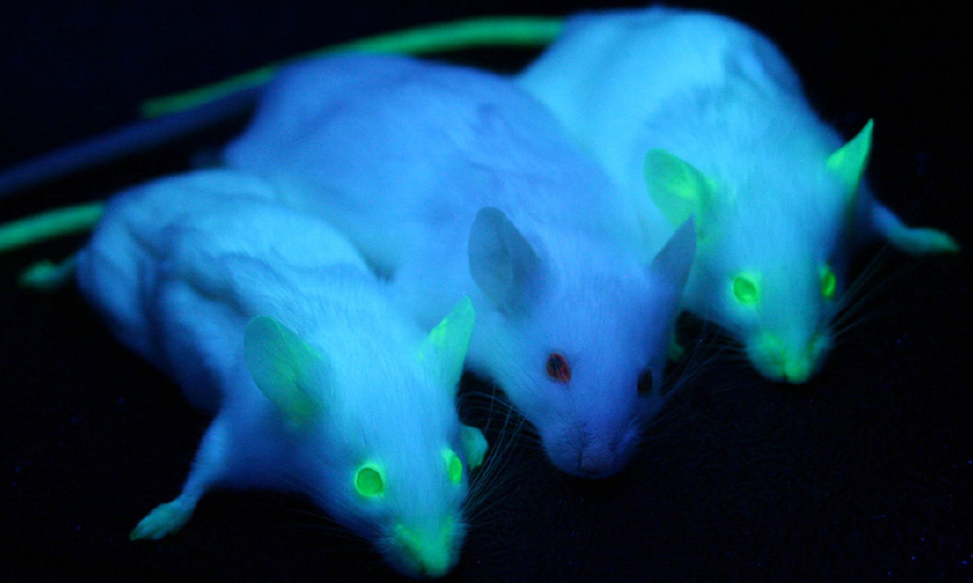
image of transgenic mice

Changes in fine structure accompanying estrogen-induced tumorigenesis of Leydig cells in the mouse testis, 1975

Abstract

The development of estrogen-induced Leydig cell tumors in cryptorchid BALB/c mice was studied with the electron microscope.

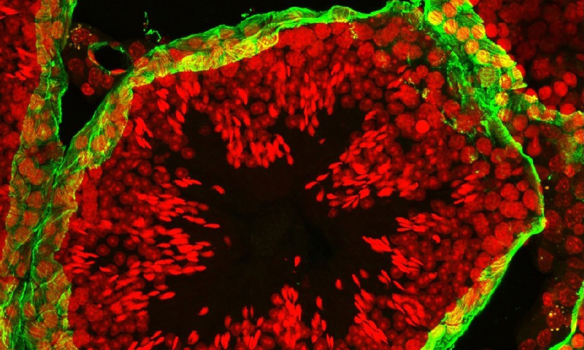
Changes in Leydig cell fine structure are apparent by 10 days after the s.c. implantation of a pellet of diethylstibestrol (DES). The smooth endoplasmic reticulum is diminished, and there is an increase in lipid droplets and free polysomes as compared with untreated cryptochid controls. These alterations persist as the Leydig cells proliferate to form focal areas of hyperplasia in the interstitial tissue. During this period of proliferation, activated macrophages containing large residual bodies appear among the Leydig cells. If DES treatment is continued for several months, malignant Leydig cell tumors, result. They are characterized by a nuclear and cytoplasmic pleomorphism of the Leydig cells and a decreased macrophage population. Virus-like particles are rarely seen within the cell during the period of tumorigenesis. Along with the reduction in smooth endoplasmic reticulum in the Leydig cells after DES treatment, evidence from the literature suggests that there is also a decrease in testosterone biosynthesis. However, it is not clear whether these two effect are correlated, since the level of the microsomal enzymes of steroid biosynthesis may vary independently of either the amount of smooth endoplasmic reticulum or the level of androgen secretion. The increase in lipid droplets seen in Leydig cells after DES treatment suggest the accumulation of precursors from the steroid biosynthetic pathway. The macrophages are though to represent scavenger cells, rather than a primary tumor cell population. The paucity of virus-like particles within altered Leydig cells implies that formed virus is not a prerequisite for tumorigenesis.

References

  • Changes in fine structure accompanying estrogen-induced tumorigenesis of Leydig cells in the mouse testis, Cancer research, NCBI PubMed, PMID: 236824, 1975.
  • Featured image credit wikimedia.
DES DIETHYLSTILBESTROL RESOURCES

Genital-tract cancers in adolescents and young adults

Stilbesterol exposure possibly linked to cancers of the testis in the 20+ year old

1972 Study Abstract

In a review of the California Tumor Registry from 1950-1969, which records 1/3 of all cancer cases in California,

  • there was an increase in the number of cancers of the vagina, corpus uteri, prostate, testis, and bladder (male) for the 10-19 year old age groupThere was no increase in cancers of the vulva, ovary, cervix, breast, stomach, colon, rectum, and bladder (female).
  • However, for the 20-24 year old age group, cancer of the vulva and testis increased from 1962-1969.

The increases are compatible with other observations which indicate a possible association between stilbesterol and other cervical sites besides the vagina. There is a need for cancer surveillance to determine disease patterns and stimulate examination of existing data.

References

DES DIETHYLSTILBESTROL RESOURCES

Testicular Leydig cell tumourigenesis by diethylstilbestrol in the BALB/c mouse

Histologic and histochemical study, European Journal of Cancer, 1966

Abstract

The present study is dealing with an investigation on gross, histologic and histochemical changes in testes and adrenal cortex of BALB/c mice after subcutaneous implantation of a diethylstilbestrol pellet. The animals were serially killed 30, 60, 90, 120, 150 and 180 days after the implantation. They showed an initial marked loss of body weight, an increased testicular weight starting from the 90th day and a constant adrenal weight increase from the beginning of the experiment. Leydig cell hyperplasia was observed in the treated groups starting from the 60th day, and Leydig cell tumours from the 120th day. A marked regression of the spermatogenic elements was observed shortly after implantation; subsequently a partial revival of the seminal activity with final regression of spermiogenesis was found. The seminal vesicles-coagulating gland complex showed a persistent squamous metaplasia of the coagulation epithelium and evidence of initial atrophy of the seminal vesicles epithelium showing a functional revival in the later stages of the experiment.

The histochemical study carried out on testes and adrenal cortex gave useful information on some enzyme activities connected with steroidogenesis, i.e. 3β-hydroxysteroid dehydrogenase, glucose-6-phosphate dehydrogenase and NADPH diaphorase. The demonstration of the reported enzymes was obtained within normal, hyperplastic and tumoural Leydig tissues. A lack of enzyme activities was found in the inner zones of the adrenal cortex as well as in some Leydig cells where ceroid deposition was frequently found.

References

  • Testicular Leydig cell tumourigenesis by diethylstilbestrol in the BALB/c mouse: Histologic and histochemical study, Histologic and histochemical study, European Journal of Cancer, doi.org/10.1016/0014-2964(66)90056-9, 1966.
  • Featured image credit bbc news health.
DES DIETHYLSTILBESTROL RESOURCES

Testicular Dysgenesis Syndrome and Testicular Germ Cell Cancer

Endocrine-Disrupting Chemicals: An Endocrine Society Scientific Statement, 2009

Abstracts

As discussed below, development of vaginal adenocarcinoma in women exposed fetally to DES and the association of carcinoma in situ in the fetal testis with the development of testicular cancer in adulthood (see Testicular dysgenesis syndrome: possible role of endocrine disrupters, 2006, and Pathways of endocrine disruption during male sexual differentiation and masculinization, 2006) are examples of links between the fetal environment and the occurrence of adult disease.

In the male, cryptorchidism, hypospadias, oligospermia, and testicular cancer have been proposed to be linked as the testicular dysgenesis syndrome (TDS) arising from disturbed prenatal testicular development. Such links are important because they could mean that several disorders occur at different periods throughout life in a single individual as a result of exposure to a given EDC (or mixture) at a particular period.

Testicular Dysgenesis Syndrome TDS

Skakkebaek et al. hypothesized that diminished semen quality, TGCC, and male urogenital tract anomalies may share a common causal pathway. They defined this triad as the TDS. The hypothesis invokes a common pathway by which EDCs, and other environmental chemicals and genetic factors, may lead to abnormal development of the fetal testis, producing testicular dysgenesis that can manifest as an increased risk of urogenital abnormalities in newborn males, as well as altered semen quality and TGCC in young men. As a cautionary note, the manifestations (or symptoms) of TDS have other causes apart from testicular dysgenesis.

It is hypothesized that TDS is due to prenatal Leydig and Sertoli cell dysfunction with secondary androgen insufficiency and impaired germ cell development. This should not be confused with the clinical diagnosis of dysgenetic testes, which is associated with genital ambiguity and a high risk of testicular malignancy. The existence of TDS as a distinct clinical entity and of possible associations with EDCs is an area of active research

Testicular Germ Cell Cancer TGCC

The earliest suggestion of epidemiological evidence related to prenatal estrogen exposure and increased risk of TGCC came from a (DES) study in 1979. However, other studies have not consistently confirmed these earlier results.

References

  • Full study (free access) : Endocrine-Disrupting Chemicals: An Endocrine Society Scientific Statement, Endocrine Reviews, PMC2726844, 2009 June.
  • Featured image credit tamassetmanagement.
DES DIETHYLSTILBESTROL RESOURCES

Pre-natal and peri-natal exposures and risk of testicular germ-cell cancer

Exposure to maternal hormones (during pregnancy), particularly estrogen (DES), is associated with testicular germ‐cell cancer risk, 2000

Abstracts

The present case-control study was undertaken to investigate the association between exposure to maternal hormones and risk of testicular germ-cell cancer (TGCC) by histologic subgroups.

Cases were males, aged 16 to 59 years, diagnosed with testicular germ-cell cancer in Ontario between 1987 and 1989.

Histologic review was performed on all eligible cases for the purpose of categorizing cases as seminoma or non-seminoma (the latter classified 2 ways, with and without tumors containing seminoma). Risk factor data were collected on 502 cases, 346 case mothers, 975 age-matched controls, and 522 control mothers.

Exogenous hormone exposure was associated with elevated risk (OR = 4.9, 95% CI 1.7-13.9). Several additional risk factors were associated with risk of testicular cancer: bleeding and threatened miscarriage (OR = 0.6, 95% CI 0.3-1.0), maternal cigarette smoking (12+ cigarettes/day OR = 0.6, 95% CI 0. 4-1.0). pre-term birth (OR = 1.6, 95% CI 1.0-2.5), and treatment for undescended testicle (OR = 8.0, 95% CI 3.2-20.0). First births were associated with elevated risk (OR = 1.7, 95% CI 1.0-2.8) among mothers below the age of 24 years at conception. There was little evidence that risk factors differed by histologic subgroup. We found evidence that exposure to maternal hormones, particularly estrogens, is associated with testicular germ-cell cancer risk. Not only does exposure to elevated levels (exogenous hormone use, pre-term birth, and first births among young mothers) increase risk but also exposure to relatively lower levels (heavy cigarette consumption and, perhaps, bleeding and threatened miscarriage) may decrease cancer risk.

Introduction

The early age at incidence of testicular germ‐cell cancer suggests that childhood exposures, possibly even those occurring in utero, may be important, including exposure to maternal hormones, from exogenous and endogenous sources.

A number of case‐control studies have interpreted results based on the hypothesis that exposure to maternal hormones elevates risk of testicular cancer (see 1980 study, 1983 study, 1986 study)  however, their results have not been entirely consistent with this hypothesis.

The present case‐control study was undertaken to examine the association of maternal hormone exposure, from both exogenous and endogenous sources, and risk of testicular germ‐cell cancer. The study also included histological review of all eligible cases, to further investigate the association according to the major histological subgroups, seminoma and non‐seminoma (the latter classified 2 ways, with and without tumors containing seminoma).

Definition of exposure variables

Exogenous hormone exposure was determined from the mother’s reported use of prescription hormones [e.g., diethylstilbestrol (DES) or premarin], prescription medication for conditions associated with threatened miscarriage, injections or pills to determine pregnancy, and use of oral contraceptives around the time of conception. Because reported exposures to exogenous hormones were uncommon, these exposures were combined into a dichotomous variable, coded “yes” if the mother reported exposure to any of the sources of exogenous hormones and “no” if she reported exposure to none of them.

Discussion

Early population‐based case‐control studies reported that exogenous hormone exposure, in the form of pregnancy tests and hormone use for threatened miscarriage, elevated risk for testicular cancer (see 1979 study, 1980 study, 1983 study), while later studies failed to confirm the association (see 1986 study, 1988 study). Despite the fact that the present study provides evidence of an association in both histological subgroups, caution should be used in interpreting the results: DES has been associated with adverse health effects in both male and female offspring, exposure status was not validated in the current study, and participation rates among case mothers were higher than among control mothers. Potential recall bias has been reported in other studies, and such bias could be expected to operate in both histological subgroups.

Conclusion

In conclusion, our results support the hypothesis that exposure to maternal hormones, particularly estrogen, is associated with testicular germ‐cell cancer risk. Not only does exposure to elevated levels of maternal hormones (exogenous hormone use and pre‐term birth) appear to increase cancer risk, but exposure to relatively lower levels of maternal hormones (heavy cigarette consumption and, perhaps, bleeding and threatened miscarriage), as measured by indices of exposure to maternal hormones, appears to decrease cancer risk. Future studies should consider direct measurement of these exposures, to further explore the association between maternal estrogen exposure and risk of testicular germ‐cell cancer.

Evidence that risk factors differ by histological subgroup has been difficult to interpret. Our results support the conclusion of Møller and Skakkebæk that the causes of seminoma and non‐seminoma are likely the same. Furthermore, there is little convincing evidence to support the need to distinguish between non‐seminomatous tumors containing seminoma and those not containing seminoma. However, caution should be used in interpreting these results because small numbers in the histological subgroup analysis resulted in limited statistical power to detect differences. Since the evidence for possible differences comes from studies looking at maternal age and parity, an understanding of the mechanisms underlying histological differences may come with a better understanding of how maternal age and parity themselves are associated with risk of testicular germ‐cell cancer.

References

DES DIETHYLSTILBESTROL RESOURCES