Prenatal DES treatment and subsequent development of testicular tumors

Lesions of the rete testis in mice exposed prenatally to diethylstilbestrol, 1985

Abstract

Adenocarcinoma of the rete testis is an exceptionally rare and malignant testicular neoplasm.

Although treatment of pregnant women with diethylstilbestrol (DES) results in reproductive tract abnormalities in their male offspring, increased incidence of testicular tumors has not been verified. However, recently three cases of seminoma have been described in men prenatally exposed to DES, suggesting an association of prenatal DES treatment and the subsequent development of testicular tumors.

This report describes the treatment of outbred pregnant CD-1 mice with DES (100 micrograms/kg) on Days 9 through 16 of gestation and its effects on their male offspring.

In addition to nonmalignant abnormalities such as retained testes which have been reported in men exposed prenatally to DES, lesions resembling adenocarcinoma of the rete testis were seen in prenatally DES-treated mice at 10 to 18 mo of age (11 of 233; 5%). No comparable lesions were seen in 96 age-matched control male mice.

These results suggest an association of prenatal DES exposure and the subsequent development of testicular lesions in the rete testis of mice.

References

  • Lesions of the rete testis in mice exposed prenatally to diethylstilbestrol, Cancer research, NCBI PubMed PMID: 4027990, 1985 Oct.
  • Featured image credit the guardian.
DES DIETHYLSTILBESTROL RESOURCES

Testicular dysgenesis syndrome

Possible role of endocrine disrupters, 2006

Abstracts

The testicular dysgenesis syndrome (TDS) hypothesis proposes that the four conditions cryptorchidism, hypospadias, impaired spermatogenesis and testis cancer may all be manifestations of disturbed prenatal testicular development. The TDS hypothesis is based on epidemiological, clinical and molecular studies, all suggestive of an interrelation between the different symptoms. The aetiology of TDS is suspected to be related to genetic and/or environmental factors, including endocrine disrupters.

…Important insight into the effect of in utero exposure to compounds with oestrogenic activity in humans comes from the former widespread use of diethylstilboestrol (DES), a synthetic oestrogen. …
… The effect of DES on sperm concentration and risk of development of testis cancer is unclear, as some studies have found a reduced sperm concentration in men exposed to DES in utero, whereas others have not. For testis cancer, only a slight increase in risk has been cautiously suggested (1996), (2001). …

Few human studies have found associations/correlations between endocrine disrupters, including phthalates, and the different TDS components. However, for ethical reasons, evidence of a causal relationship between prenatal exposure and TDS is inherently difficult to establish in human studies, rendering the recently developed animal TDS model an important tool for investigating the pathogenesis of TDS. Clinically, the most common manifestation of TDS is probably a reduced sperm concentration, whereas the more severe form may include a high risk of testis cancer. Clinicians should be aware of the interconnection between the different features of TDS, and inclusion of a programme for early detection of testis cancer in the management of infertile men with poor semen quality is recommended.

References

  • Testicular dysgenesis syndrome: possible role of endocrine disrupters, Best practice & research. Clinical endocrinology & metabolism, NCBI PubMed PMID: 16522521, 2006 Mar.
DES DIETHYLSTILBESTROL RESOURCES

Cancer risk in men exposed in utero to diethylstilbestrol

DES Sons’ (increased) cancer risk, 2001

Abstracts

BACKGROUND
An association between prenatal diethylstilbestrol (DES) exposure and cancer in men, especially testicular cancer, has been suspected, but findings from case-control studies have been inconsistent. This study was conducted to investigate the association between prenatal DES exposure and cancer risk in men via prospective follow-up.

METHODS
A total of 3613 men whose prenatal DES exposure status was known were followed from 1978 through 1994. The overall and site-specific cancer incidence rates among the DES-exposed men were compared with those of the unexposed men in the study and with population-based rates. The relative rate (RR) was used to assess the strength of the association between prenatal DES exposure and cancer development. All statistical tests were two-sided.

RESULTS
Overall cancer rates among DES-exposed men were similar to those among unexposed men (RR = 1.07; 95% confidence interval [CI] = 0.58 to 1.96) and to national rates (RR = 0.99; 95% CI = 0.65 to 1.44). Testicular cancer may be elevated among DES-exposed men, since the RRs for testicular cancer were 3.05 (95% CI = 0.65 to 22.0) times those of unexposed men in the study and 2.04 (95% CI = 0.82 to 4.20) times those of males in the population-based rates. The higher rate of testicular cancer in the DES-exposed men is, however, also compatible with a chance observation.

CONCLUSIONS
To date, men exposed to DES in utero do not appear to have an increased risk of most cancers. It remains uncertain, however, whether prenatal DES exposure is associated with testicular cancer.

References

  • Cancer risk in men exposed in utero to diethylstilbestrol, Journal of the National Cancer Institute, NCBI PubMed PMID: 11287449, 2001 Apr.
  • Featured image Harshil Gudka.
DES DIETHYLSTILBESTROL RESOURCES

DES-induced Testis Cancer

Male reproductive health and environmental xenoestrogens, 1996

Abstracts

Incidence of Testicular Cancer

Testicular cancer is now the most common malignancy of young men in many countries; and although it is still rare compared to the malignant diseases most prevalent in old age, the lifetime risk of developing testicular cancer now approaches 1% in a country such as Denmark. The incidence of testicular cancer has increased for several decades.

Risk factors for testicular cancer have been analyzed in several case-control studies. Estrogen treatment of mothers whose sons have developed testicular cancer has remained an equivocal risk factor. It is unfortunate that there are no prospective studies yet concerning the testicular cancer risk among males exposed to DES.

The effect of DES on risk of development of testis cancer seems to be unclear; only a slight increase in risk has been cautiously suggested.

Occurrence of Abnormalities in the Reproductive System of the Sons of Women Exposed to Diethylstilbestrol during Pregnancy

Summary

Exposure to DES during pregnancy results in an increased risk for several male reproductive disorders, such as cryptorchidism, urethral abnormalities, epididymal cysts, and testicular hypoplasia. In addition, the semen quality of DES sons is worse than that of controls. Incidence of testicular cancer is approximately doubled among DES sons compared to the general population, but whether this represents a true increase of the cancer risk is equivocal.

Effects of Synthetic Estrogens on the Testis in Animal Models

Summary

Diethylstilbestrol treatment of experimental animals in utero results in increased incidence of cryptorchidism; urethral abnormalities; testicular hypoplasia; poor semen quality; and infertility, abnormalities in accessory sex organs, rete testis adenocarcinoma, interstitial cell hyperplasia, and tumors. Thus, the outcome of DES exposure of experimental animals is highly analogous to the findings in humans.

References

  • Full study (free access) : Male reproductive health and environmental xenoestrogens, Environmental Health Perspectives, NCBI PubMed PMC1469672, 1996 Aug.
  • Featured image clevelandclinic.
DES DIETHYLSTILBESTROL RESOURCES

DES immunotoxicity

The comparative immunotoxicity of five selected compounds following developmental or adult exposure, 2006

Study Abstract

It is well established that human diseases associated with abnormal immune function, including some common infectious diseases and asthma, are considerably more prevalent at younger ages. Although not established absolutely, it is generally believed that development constitutes a period of increased immune system susceptibility to xenobiotics, since adverse effects may occur at lower doses and/or immunomodulation may be more persistent, thus increasing the relative risk of xenobiotic exposure to the immunologically immature organism.

To address this issue, a brief overview of immune maturation in humans is provided to demonstrate that functional immaturity alone predisposes the young to infection. Age-dependent differences in the immunotoxic effects of five diverse compounds, diethylstilbestrol (DES), diazepam (DZP), lead (Pb), 2,3,7,8-tetrachlorodibenzo-p-dioxin (TCDD), and tributyltin oxide (TBTO), which have undergone adult and developmental immunotoxicity testing in rodents, are then reviewed, as are human data when available. For all five chemicals, the developing immune system was found to be at greater risk than that of the adult, either because lower doses produced immunotoxicity, adverse effects were more persistent, or both.

Diethylstilbestrol immunotoxicity

Background

Between 5 and 10 million pregnant women were given diethylstilbestrol (DES), a potent synthetic nonsteroidal estrogen, between 1938 and 1971 to prevent premature delivery or pregnancy loss. Its use was terminated when a rare form of reproductive system cancer was found in female offspring of DES-exposed mothers. Male and female reproductive systems malformations have been reported in children of treated women, as has anecdotal evidence of immune system dysfunction. DES was also used to increase weight gain in livestock, although this use is no longer permitted in most countries.

Effects on the Immune System in Humans

Both female and male children of DES-exposed mothers report a higher incidence of autoimmune diseases and asthma (Baird et al., 1996). In general, these diseases are considered to be the result of inappropriate immune system responses, or possible loss of homeostatic control, instead of immune system suppression.

Effects on the Immune System in Rodents

In utero exposure
Luster et al. (1978b) reported that a single injection of 0.1 mg DES/kg body weight on gestational day (GD) 16 did not affect the antibody response to the T-cell-dependent antigen, sheep red blood cells (SRBC), when evaluated in 7-wk-old male and female offspring of Swiss-Webster mice. The T-independent IgM response of female offspring to bacterial lipopolysaccharide (LPS) was suppressed by DES, but was similar to control responses when females were
immunized for a second time. In marked contrast, the male offspring response to LPS immunization was enhanced after both first and second immunizations, an effect attributed to the stimulating effect of estrogen on the antibody response to LPS. Delayed-type hypersensitivity responses (DTH) were suppressed in female, but not in male, offspring, even though thymus weights and T-cell responses to polyclonal stimulation were suppressed in both genders (Luster et al., 1979). Further studies suggest that DES targets early precursors of T lymphocytes in the fetal liver, accounting for thymic atrophy and suppression of DTH (Holladay et al., 1993), but not for defects in T-independent responses to LPS of female offspring.

Neonatal exposure
Nonspecific T- and B-cell proliferation was reported to be suppressed in 6-wk-old female NMRI mice given 5 μg DES/d (roughly 2.2 mg DES/kg/d) over postnatal days (PND) 1–5 (Kalland et al., 1979); suppression was still evident at 17 mo of age (normal life span ~24 mo). It is noteworthy that neither estradiol nor corticosterone exposure over PND 1–5 produced long-term suppression, and that lymphocyte proliferation was comparable to control values at 6 wk of age in females exposed to DES over PND 6–10. Lower doses (approximately 4.4, 44, or 440 μg/kg/ d) had no effect on proliferative response. The 5-μg DES/d exposure regimen also decreased NK cell activity in 6- to 8-wk-old female inbred C57Bl/6 (75%↓) and BALB/c (53%↓) mice and in outbred NMRI (28%↓) mice (Kalland, 1980a). NMRI or AKR/J female mice, exposed to 5 μg DES/d over PND 1–5, were also more likely to develop tumors after low dose injection of a known carcinogen (Kalland & Forsberg, 1981). A subsequent paper (Kalland, 1984) reported that, on a per cell basis, NK cells from DES mice were as active as cells from the control group, but that exposure reduced the number of NK cell precursors in the bone marrow. In other words, NK cells from experimental animals were as efficient as those from controls, but a deficiency in NK cell precursors produced functional suppression of NK activity at the whole animal level. The same postnatal exposure regimen (Kalland, 1980a) reduced the T-lymphocyte-dependent antibody response to SRBC by ~60%, and the T-independent response to bacterial LPS by ~40% when examined in 16- to 18-wk-old NMRI mice. Suppression of the T-dependent response was reportedly due to a defect in T-helper cells. DTH responses were likewise suppressed in 6- and 9-mo-old NMRI females exposed to approximately 2.2 mg/kg/d over PND 1–5 (Kalland & Forsberg, 1978). Kalland (1980b) also reported a persistent (at least 6.5 mo postpartum) decrease in the proportion of T cells in the spleens of DES-exposed mice.

Adult exposure
Luster et al. (1980) reported suppression of the antibody response to SRBC or LPS, and the DTH to keyhole limpet hemocyanin (KLH), in adult female mice exposed to 2 or 8 mg DES/kg/d × 5 d. The DTH was decreased in mice dosed with DES after, but not before, sensitization with KLH, suggesting that the suppressive effects of DES on DTH were not persistent. Using the same exposure regimen, resistance to bacterial or parasite infection was decreased and tumor incidence in animals challenged with tumor cells was increased at ≥2 mg DES/kg/d (Dean et al., 1980). T-cell-mediated resistance to a nematode infection was suppressed by 5 d of exposure to 0.2 mg DES/kg/d if exposure began on the day of infection; if exposure commenced 5 d before or 3 or 8 d after infection, decreased resistance was only observed at the highest dose (8 mg/kg/d) (Luebke et al., 1984).

Mode(s) of Action

DES is a potent estrogen, and likely affects immune function via the estrogen receptor (ER). Evidence includes similar effects of known estrogens (17β-estradiol) on the immune system of adult and neonatal rodents, blockade of certain immunotoxic effects by pharmacologic antagonism of the ER (Luster et al., 1984), and antagonism of estrogen-mediated immune system effects in mice lacking ERα (Staples et al., 1999). DES appears to target precursor cells in the bone marrow (adults and neonates) and fetal liver (neonates), producing a long-lasting or perhaps permanent reduction in numbers of precursor cells. This defect explains a significant portion of long-lived immunosuppressive effects (e.g., Kalland’s 1984 paper on suppressed NK activity), although the effects of adult exposure also includes damage to the thymic epithelium (Luster et al., 1984). The underlying mechanism of long-term suppression following exposure of the developing immune system to DES is not known, but the default assumption is that a critical cell population is lost to developmental exposure; either this purported population is refractory to estrogen-mediated ablation in adults or repair and recovery mechanisms are present in adults that are lacking in the developing immune system.

Data Gaps

There has been no systematic evaluation of persistent DES-mediated immunosuppression in adult animals. Dose-response data are not available for many of the of the developmental exposure studies that revealed persistent effects.

Summary

In utero exposure to 0.1 mg DES/kg during the last trimester of pregnancy suppressed T-cell- and B-cell-mediated responses only in female offspring. The gender dependence of effects was remarkable in that T-independent responses in male offspring were enhanced, yet suppressed in females. Exposure during gestation produced effects that persisted into the equivalent of young adulthood. In neonates there appears to be a critical developmental window during PND 1–5, during which exposure to DES produces persistent immune system defects that last well into adulthood or persist for most of the normal life span of the mouse. These effects are among the most persistent reported for any chemical. In adults, immunosuppression occurs at doses similar to those that produce immunotoxicity in developing animals. However, the immune system-related endpoints that have been evaluated over time in exposed adult animals (bone marrow cellularity, thymus weights) recover relatively quickly (Forsberg, 1984). In adults, recovery may occur so quickly that suppression of cell function or resistance to infection may require ongoing exposure to maintain suppression.

Conclusions

Immunotoxicity has been reported at similar doses when exposure occurs during late gestation, early postpartum, or as adults. However, the distinguishing feature of developmental exposure to DES is the persistence of effects, some of which are still apparent in very old mice. In contrast, immune system-related endpoints that have been evaluated (bone marrow cellularity, thymus, weights) suggest that adults recover relatively quickly (Forsberg, 1984).

References

  • The comparative immunotoxicity of five selected compounds following developmental or adult exposure, Journal of toxicology and environmental health. Part B, Critical reviews, NCBI PubMed, PMID: 16393867, 2006 Jan-Feb.
  • Featured image radicalremission.
DES DIETHYLSTILBESTROL RESOURCES

Prenatal DES exposure linked to Hashimotos thyroiditis

Drug exposure, pregnancy outcome and fetal and childhood development occurring in the offspring of mothers with systemic lupus erythematosus and other chronic autoimmune diseases

2006 Study Abstract

Most autoimmune diseases occur more commonly in females and many of these young women wish to become mothers. For pregnancy to proceed successfully immunomodulation and physiological changes preparing the reproductive system need to occur.

Pregnancy occurring in a chronically ill mother who requires medications in order to maintain her own health and who may have already incurred significant organ pathology gives rise to several problems and so four questions arise:

  1. What will be the effect of the pregnancy on the underlying disease?
  2. What will be the effect of the disease on the outcome of pregnancy?
  3. How to manage the disease, just prior to, throughout and immediately after the pregnancy?
  4. The long term fetal and childhood effects of maternal disease and its management.

This paper reviews the current literature pertaining to these questions in patients with systemic lupus erythematosus (SLE) and other chronic rheumatic and autoimmune diseases.

Diethylstilbestrol

… “Other evidence suggests that the risk of autoimmune disease, particularly Hashimotos thyroiditis, is also increased in individuals exposed to DES in utero.” …

References

  • Drug exposure, pregnancy outcome and fetal and childhood development occurring in the offspring of mothers with systemic lupus erythematosus and other chronic autoimmune diseases, Lupus, NCBI PubMed, PMID: 17153855, 2006.
  • Featured image cleveland clinic.
DES DIETHYLSTILBESTROL RESOURCES
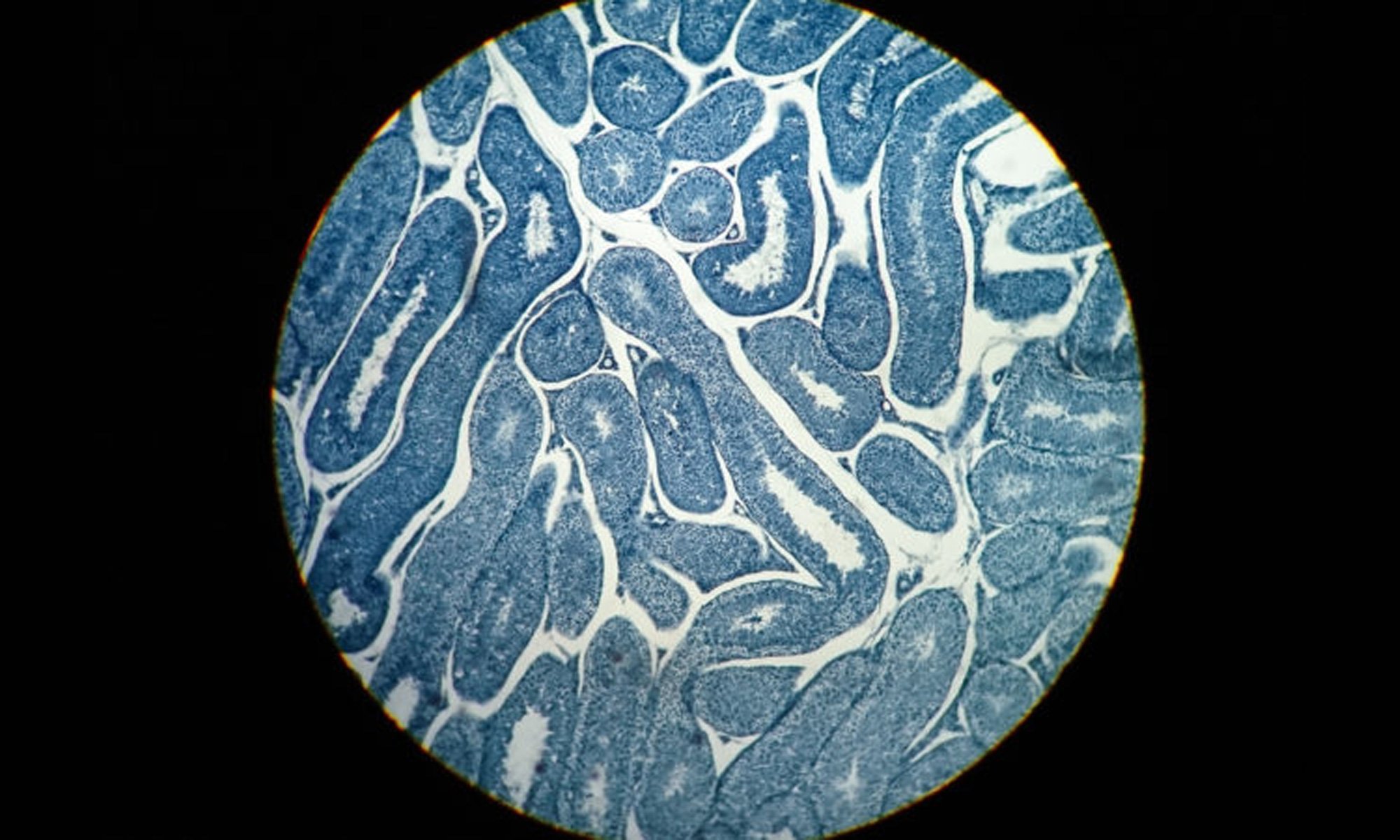
How DES exposure leads to reduced fertility in males

Diethylstilbestrol induces morphological changes in the spermatogonia, Sertoli cells and Leydig cells of adult rat

2019 Study Abstract

It is now established that diethylstilbestrol (DES) has damaging effects on the male reproductive system. However, to date there have been no studies morphological analysis of adult rat testes upon treatment with DES.

Here, we examined whether DES has any significant morphological effect on steroidogenesis and spermatogenesis.

DES was injected subcutaneously at 3 μg/day and 30 μg/day in adult male Sprague-Dawley (SD) rats for two different treatment lengths (1 or 3 weeks), after which rats were necropsied. TUNEL labeling, cell counting, and morphological analysis were used to evaluate the effects of DES.

A high dose of DES and longer exposure severely affected the cellular development of the testis. Specifically, DES treatment disrupted both steroidogenesis and spermatogenesis by decreasing the number of spermatogonia, Sertoli cells, and Leydig cells in a dose- and time-dependent manner.

Thus, DES may account for decreases in the number of spermatogenic cells, Sertoli cells and Leydig cells, which in turn may lead to reduced fertility in males.

References

  • Diethylstilbestrol induces morphological changes in the spermatogonia, Sertoli cells and Leydig cells of adult rat, Research in veterinary science, NCBI PubMed, PMID: 31082573, 2019 Apr.
  • Featured image Bart Plantenga.
DES DIETHYLSTILBESTROL RESOURCES
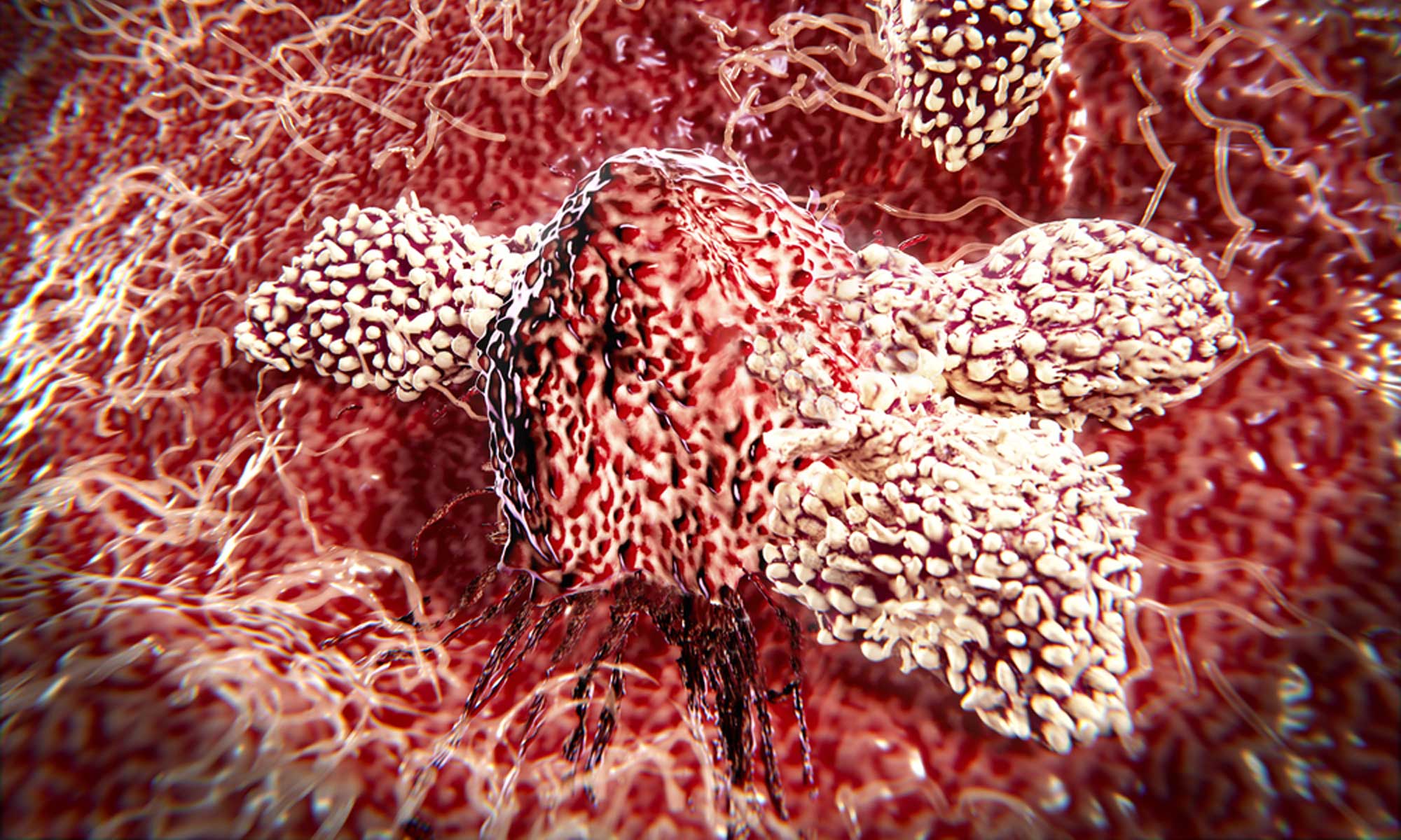
DES exposure causes decreased testis weight and morphological demasculinization of males

Short-term study investigating the estrogenic potency of diethylstilbesterol in the fathead minnow (Pimephales promelas)

2012 Study Abstract

Diethylstilbestrol (DES) is a synthetic estrogen that has been “banned” for use in humans, but still is employed in livestock and aquaculture operations in some parts of the world.

Detectable concentrations of DES in effluent and surface waters have been reported to range from slightly below 1 to greater than 10 ng/L. Little is known, however, concerning the toxicological potency of DES in fish.

In this study, sexually mature fathead minnows (Pimephales promelas) of both sexes were exposed to 1, 10, or 100 ng of DES/L of water in a flow-through system. Tissue concentrations of DES and changes in a number of estrogen-responsive end points were measured in the fish at the end of a 4 d exposure and after a 4 d depuration/recovery period in clean water. Accumulation of DES was sex-dependent, with females exhibiting higher tissue residues than males after the 4 d exposure. The observed bioconcentration of DES in the fish was about 1 order of magnitude lower than that predicted on the basis of the octanol-water partition coefficient of the chemical, suggesting relatively efficient metabolic clearance by the fish. Exposure to 1, 10, or 100 ng of DES/L caused decreased testis weight and morphological demasculinization of males (regression of dorsal nuptial tubercles). Diethylstilbesterol induced plasma vitellogenin (VTG) in both sexes at water concentrations ≥10 ng/L; this response (especially in males) persisted through the end of the 4 d recovery period. Hepatic transcripts of VTG and estrogen receptor-α also were affected at DES concentrations ≥10 ng/L. Evaluation of transcript profiles in the liver of females using a 15K-gene fathead minnow microarray revealed a concentration-dependent change in gene expression, with mostly up-regulated transcripts after the exposure and substantial numbers of down-regulated gene products after depuration. Genes previously identified as vitellogenesis-related and regulated by 17β-estradiol were significantly enriched among those differentially expressed following exposure to DES.

Overall, our studies show that DES causes a range of responses in fish at water concentrations comparable to those reported in the environment and that in vivo potency of the estrogen is on par with that of the better-studied estrogenic contaminant 17α-ethinylestradiol.

References

  • Short-term study investigating the estrogenic potency of diethylstilbesterol in the fathead minnow (Pimephales promelas), Environmental science & technology, NCBI PubMed, PMID: 22708615, 2012 Jul.
  • Featured image situbiosciences.
DES DIETHYLSTILBESTROL RESOURCES
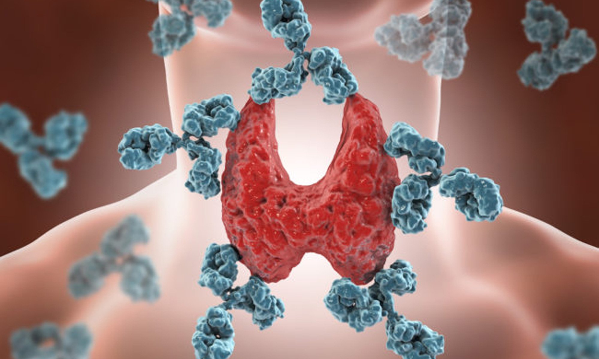
Antenatal exposure to DES: lessons learned…future concerns

DES-exposed offspring : certain complications have no time limit and continued follow-up is necessary, 2007

Summary

The short- and long-term effects of the widespread use of diethylstilbestrol (DES) over 3 decades have become a distant memory for many clinicians. Others are too young to remember the flurry of activity in the early 1970s on the part of many medical centers to identify the offspring of women who were prescribed DES during their pregnancies.

This medication was given in an attempt to prevent multiple pregnancy-related problems such as miscarriage, premature birth, and abnormal bleeding.

The recognition of the association of DES with an increased incidence of cervical and vaginal cancers in very young women led the Food and Drug Administration to ban its use during pregnancy in 1971.

Other pregnancy-related problems for the daughters and genitourinary tract changes in the sons did not become apparent until years later.

Ongoing follow-up of these offspring has raised concerns for their future as well as their mothers’ future. Clinicians need to be up-to-date with current knowledge regarding risks for cancer and other health-related issues.

Abstract (Third-Generation Effects)

Animal studies have shown tumor growth in older third-generation mice (human equivalent to age 70).

Multigenerational studies in humans are currently underway.

Several small studies of teenage third-generation females have not shown the same type of changes as in their mothers. Sons of DES daughters are at increased risk for hypospadias.

References

  • Antenatal exposure to DES: lessons learned…future concerns, Obstetrical and gynecological survey, NCBI PubMed PMID: 17634156, 2007 Aug.
  • Image credit wise owl tea ‏.
DES DIETHYLSTILBESTROL RESOURCES
DES and Cryptorchidism

EDC-2: The Endocrine Society’s Second Scientific Statement on Endocrine-Disrupting Chemicals, 2015

Abstract

It was subsequently determined that exposed offspring of both sexes had increased risk for multiple reproductive disorders, certain cancers, cryptorchidism (boys), and other diseases, although the risk for sons is more controversial.

New data are emerging to implicate increased disease risk in grandchildren.

Reference

  • EDC-2: The Endocrine Society’s Second Scientific Statement on Endocrine-Disrupting Chemicals, Endocrine Reviews, Volume 36, Issue 6, Pages E1–E150, doi.org/10.1210/er.2015-1010, 01 December 2015.
DES DIETHYLSTILBESTROL RESOURCES